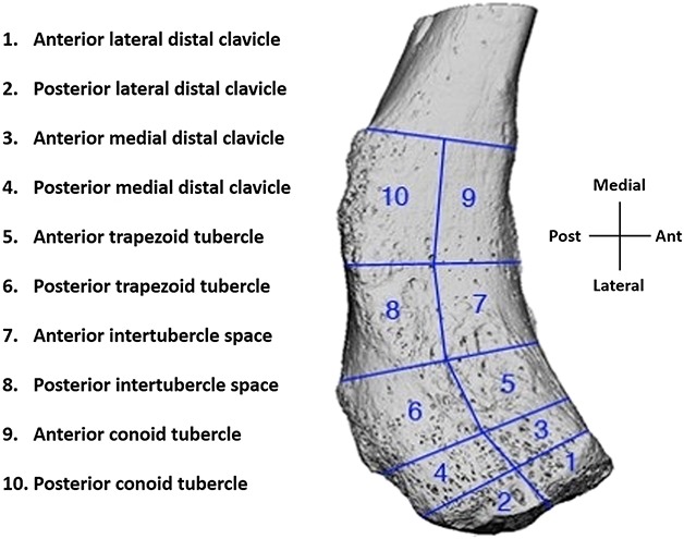
Fig. 1.

This image shows the distal clavicle regions of interest (ROI). The areas of the conoid tubercle, intertubercle space, trapezoid tubercle, and medial and distal halves of the remaining distal clavicle (distal to the trapezoid tubercle) were identified and each were divided into the anterior and posterior zones. This yielded 10 ROI that were isolated in a similar fashion in each specimen. Each anatomic ROI was assigned a number for simplicity.
