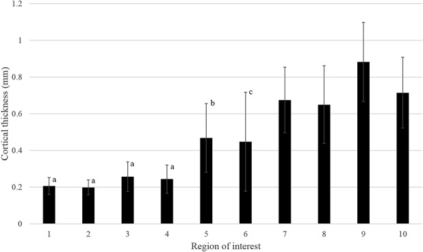
Fig. 3.

The mean cortical thickness (mm) for all regions of interest (ROI) is plotted with associated SD bars. The four most-medial ROIs demonstrated greater cortical thicknesses than the four most-lateral regions (p < 0.001 for all). Furthermore, ROI 9 and ROI 10 both demonstrated greater cortical thickness than ROI 5 (p < 0.001 for both). Also, and ROI 8 demonstrated greater cortical thickness than ROI 6 (p < 0.001). athinner cortex than ROIs 7 to 10. bthinner cortex than ROI 9 and ROI 10. cthinner cortex than ROI 8.
