
Fig. 1.
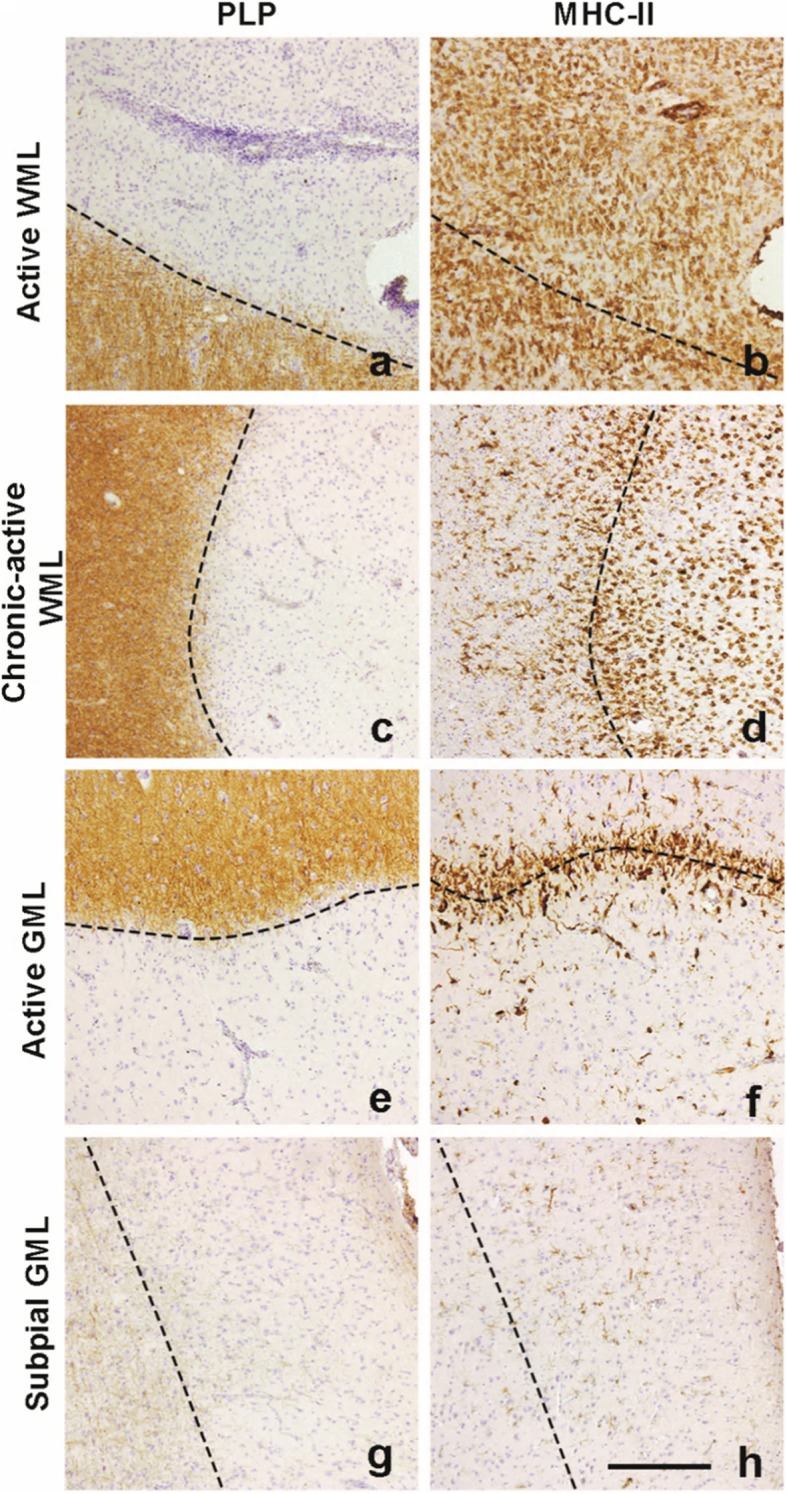
Representative images of lesion types used in this study. Lesions are characterized by loss of PLP staining and amount of MHC-II+ cells. A large amount of of MHC-II+ cells can be observed in the demyelinated area (a) in active WMLs (b). Chronic-active demyelinated WMLs (c) feature a ‘rim’ of MHC-II cells (d) which is also visible in demyelinated active GMLs (e, f). Subpial demyelinated (g) GMLs hardly show MHC-II+ cells (h). Scalebar (a-h) = 200 μm. Dashed lines indicate the edge of the lesion