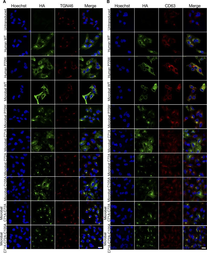
Figure S2. Subcellular localization of IFITM3 proteins and cysteine mutants of microbat IFITM3.
(A, B) Immunofluorescence of A549 cells stably expressing the indicated HA-tagged huIFITM3 or mbIFITM3 proteins (or untransduced A549 cells) were fixed, permeabilized, and stained for HA and either the trans Golgi Network marker TGN46 (A) or the endolysosomal marker CD63 (B). Nuclei were labelled with Hoechst and images taken on a confocal microscope. Scale bar represents 20 μm.

