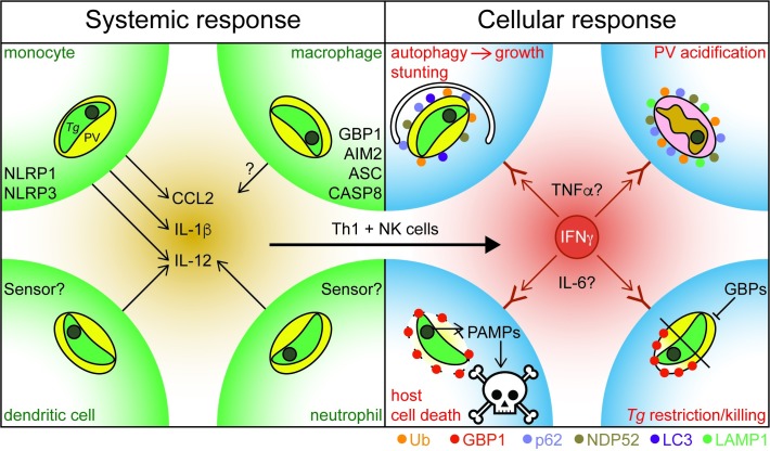
Fig 1. Systemic and cellular human response to Toxoplasma infection.
Left: Systemic response to Tg-infection: Infected cells and cells that have phagocytosed Tg parasites at a site of infection sense the presence of the pathogen via the indicated PRRs and defence proteins and react by production of proinflammatory cytokines and chemokines like CCL2, IL-1β, and IL-12. This cytokine presence will trigger IFNγ–production by Th1 and NK cells. Right: Cellular response to Tg-infection: IFNγ and potentially other cytokines trigger infected cells to mount a cell-intrinsic defence against the PV. Mechanisms include ubiquitin-driven non-canonical autophagy of the entire PV and growth stunting; marking the PV with Ub, LAMP1, and the autophagy adapter proteins NDP52 and p62, followed by acidification of the vacuoles and killing of the parasite; recruitment of GBP1 to Tg vacuoles to disrupt them and expose the parasite within or growth restriction of Tg by GBP1 without translocation to the vacuole; and host cell death in response to opened PVs and leakage of pathogen-associated molecular patterns into the cytosol for detection by PRRs. The exact mechanisms highly depend on the cell type and the Tg strain infecting the cells. AIM, absent in melanoma 2; ASC, apoptosis-associated speck-like protein containing a CARD; CASP, caspase; CCL, chemokine (C-C motif) ligand; GBP, guanylate binding protein; IL, interleukin; IFNγ, interferon gamma; LAMP, lysosome-associated membrane protein; NDP52, nuclear domain 10 protein 52; NK, natural killer; NLRP, nucleotide-binding oligomerization domain, Leucine rich repeat and Pyrin domain containing; PAMP, pathogen-associated molecular patterns; PRR, pattern recognition receptor; PV, parasitophorous vacuole; Tg, Toxoplasma gondii; Th, T helper cell; TNFα, tumour necrosis factor α; Ub, Ubiquitin.

