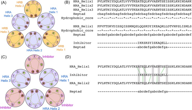
Fig 1.
A) Heptad repeat representation of the 6-HB domain formed by HRA (purple circles) and HRB domain (yellow circles). Helices are represented as circles. Amino acid heptad repeat positions are labelled with letters a through to g with hydrophobic amino acids occupying a and d positions. B) Heptad repeat assignment of HRA and HRB domain helices along with the designed inhibitor. Inhibitor heptad positions were assigned identical to the HRB domain. The hydrophobic core of residues in the a and d positions are marked with *. C) Heptad repeat representation of HRA (purple circles) domain and bound inhibitor (pink circles) replacing HRB domain. D) Salt bridges (green dotted lines) and interactions (grey bar) between the residues of the inhibitor and the HRA domain.

