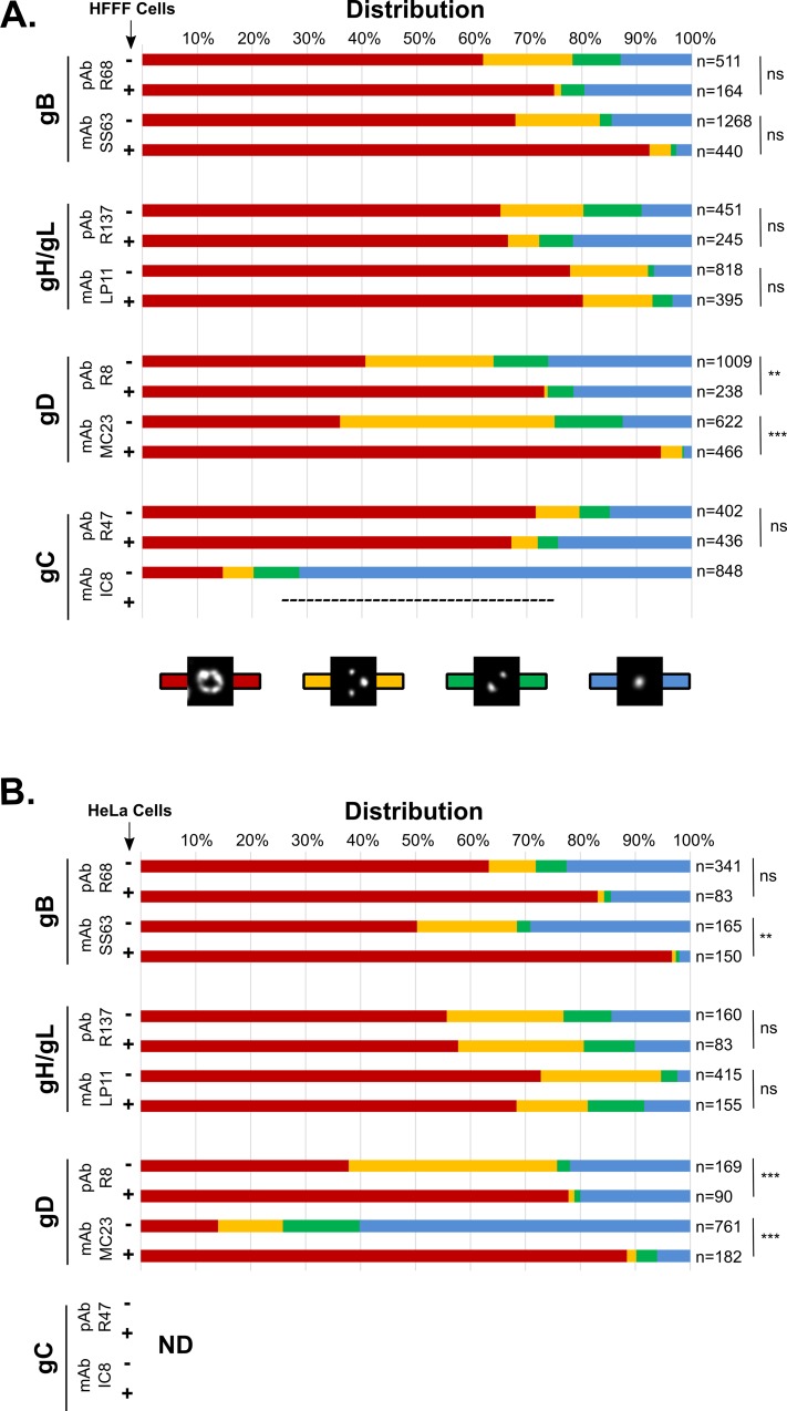
Fig 2. Free and cell-bound particles may display different glycoprotein patterns.
Particles bound to cells (+) or attached to coverslips (-),were imaged using gSTED and the localizations of glycoproteins gB, gH/gL, gD and gC were assessed by using a monoclonal (mAb) or a polyclonal antibody (pAb) recognizing each glycoprotein. Localization was categorized as single (blue), double (green) or multiple spots (yellow) or rings (red), as described in Fig 1. (A) Cells used for attachment were HFFF cells. Particles were attached to glass coverslips at room temperature whereas particles were bound to cells at 4°C to prevent membrane fusion. No IC8-positive signal (gC) could be detected on cell-bound particles. (B) Cells used for attachment were HeLa cells. Particles were attached to glass coverslips or bound to cells at 4°C. Localization of gC was not analyzed in this experiment. A Pearson’s chi-squared test was used to determine whether the profile of distribution of one glycoprotein was statistically different between free and cell-bound virions. The p-value indicates the likelihood of a correlation, therefore a p-value > 0.05 was considered as indicating a statistically significant difference between the two sets. ns: p<0.05, **: p>0.1, ***: p > 0.5. n: number of analyzed particles per condition. ND: not determined.

