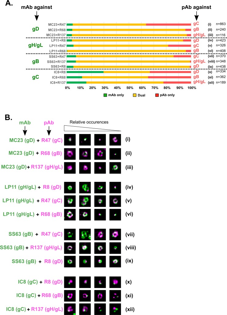
Fig 5. Relative positions of glycoproteins on free virus particles as revealed by dual-color gSTED.
(A) Purified free particles were double-labeled with twelve different pairs of antibodies. In each case, the percentage of particles labeled with mAb only is shown in green, the percentage of particles labeled with pAb only is shown in red and the percentage of double-labeled particles is shown in yellow. n: number of analyzed particles per condition. (B) For every pair of antibodies, representative noise-filtered gSTED pictures of cell-free particles are shown. The images on the left show more commonly observed patterns while those on the right are less commonly observed. mAb staining is pseudo-colored in green and pAb staining is pseudo-colored in magenta. All pictures are 700 x 700 nm.

