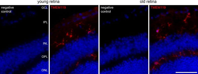
Figure 2.
Immunohistochemical labeling of microglial cells in retinal cryosections of a young and old mouse against TMEM119 (red, antibody by Synaptic Systems) as indicated. Corresponding negative controls are included. GCL, ganglion cell layer; IPL, inner plexiform layer; INL, inner nuclear layer; OPL, outer plexiform layer; ONL, outer nuclear layer. Scale bar: 50 μm.

