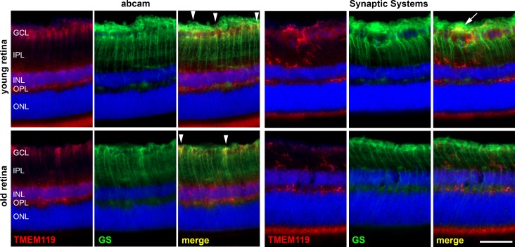
Figure 3.
Immunohistochemical staining of a retinal cryosection from a young and old mouse against TMEM119 (red) and GS as indicated. Anti-TMEM119 antibodies were delivered by Abcam (left) and by Synaptic Systems (right). With the anti-TMEM119 antibody by Abcam, a poor labeling of microglial cell was achieved, and a clear IR of other cell populations in the retina; for example, the Müller cells, as demonstrated by GS IR. White arrowheads point to particularly obvious colocalization of TMEM119 IR and GS IR. White arrow points to an overlap of TMEM119 IR and GS IR without real colocalization. Scale bar: 50 μm.

