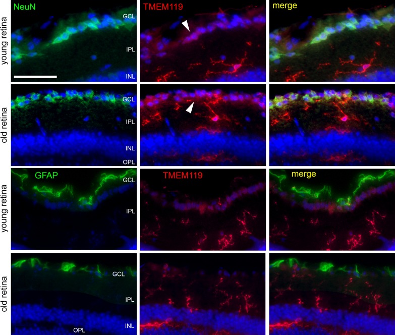
Figure 6.
Immunohistochemical staining of retinal cryosections to label microglial cells (TMEM119, antibody by Synaptic Systems, red), ganglion cells (NeuN, green), and astrocytes (GFAP, green) in retinas of young and old mice, as indicated. Only inner layers of the retinas are shown. White arrowheads point to TMEM119-positive retinal ganglion cells. Scale bar: 50 μm.

