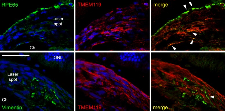
Figure 9.
Confocal images of double labeling of the proliferation area in the laser spot for TMEM119 (red, antibody by Synaptic Systems) and RPE65 (green, upper row) and vimentin (green, lower row). Colocalization could be found only in very few cases (white arrowheads). ONL, outer nuclear layer; Ch, choroid. Scale bar: 50 μm.

