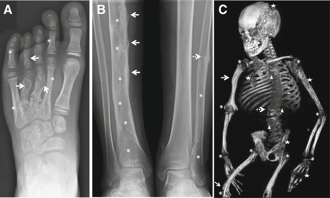
Fig. 2.
Representative radiographic skeletal features in CSHS. a Left foot radiograph (CSHS101) showing dysplastic lesions in phalanges and metacarpals of digits 2–5. In contrast, the bones of the first ray are spared from dysplasia, consistent with the mosaic nature of the syndrome. Dysplastic bone lesions are characterized by areas of mixed lysis/sclerosis. In this radiograph, the lytic component is more prominent in digits 3 and 4 (arrows), while the sclerotic component is more noticeable in digits 2 and 5 (asterisks). b Radiograph of mid and lower shafts of tibiae and fibulae (CSHS105). The right tibia shows cortical pseudofractures with periosteal reaction along the mid shaft (arrows) in addition to irregular lucent changes indicative of dysplasia (asterisks). A healing stress fracture is also observed in the mid left fibula (dashed arrow), with lucent changes in its mid and distal shaft (asterisks). The left tibia appears normal and unaffected, suggesting that dysplastic bones are more prone to fracture under the same systemic hypophosphatemic milieu than non-dysplastic bones. c Skeletal 3D CT reconstruction (CSHS106) showing severe rickets (asterisks) and lower limb bowing (arrow) secondary to osteomalacia. Patchy areas with lytic lesions are seen throughout the skeleton (stars) in addition to areas of sclerosis (compound arrow). This was the only patient in the cohort in which vertebral dysplasia was identified (triangles). Scoliosis, a frequent manifestation in CSHS, was severe in this patient (spotted arrow)

