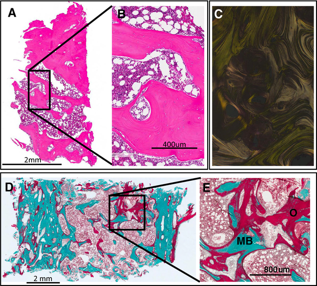
Fig. 3.
Bone histopathology. An iliac crest biopsy was performed in an area that appeared dysplastic on imaging in subject CSHS105. The same HRAS G13R mutation that had been identified in the skin was identified in bone marrow stromal cells extracted from this sample. a, b. H&E of the iliac crest does not exhibit any noticeable histopathological abnormality. c. Polarized light shows normal lamellar distribution of the collagen fibrils of the sample shown in a, b. d, e. Goldner’s trichrome stain of an undecalcified sample of the biopsy showing areas with excessive accumulation of osteoid indicative of severe osteomalacia. O (red) = osteoid; MB (green) = mineralized bone (color figure online)

