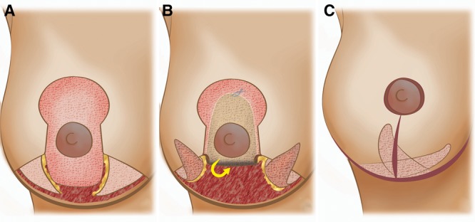
Fig. 1.

Illustration summarizing the different surgical steps. A, Superiorly based pedicled glandular flap dissected and both dermal flaps de-epidermized. B, Cranial rotation to increase upper pole fullness. C, Final result after medial fixation of the lateral triangular flap.
