Fig. 2.

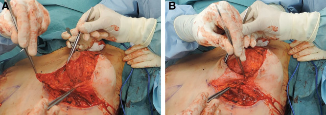
Intraoperative pictures illustrating the dermal flaps. A, Both dermal flaps are de-epidermized and glandular flap is cranially rotated. B, Repositioning of dermal flaps that will act as support.

Intraoperative pictures illustrating the dermal flaps. A, Both dermal flaps are de-epidermized and glandular flap is cranially rotated. B, Repositioning of dermal flaps that will act as support.