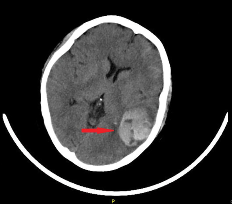
Fig. 1.

There was mixed density of left temporal lobe, indicating the amount of bleeding was around 15 ml. There was multiple high density in left frontal temporal parietal lobe and sulcus and subarachnoid hemorrhage and sulcus became shallow.

There was mixed density of left temporal lobe, indicating the amount of bleeding was around 15 ml. There was multiple high density in left frontal temporal parietal lobe and sulcus and subarachnoid hemorrhage and sulcus became shallow.