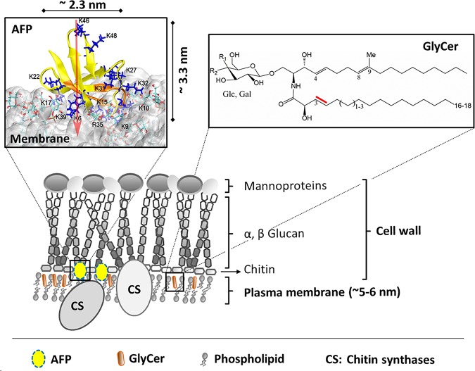
FIG 8.
Working model for the mode of action of AFP. See the text for details. Note that the upper left picture is reproduced from Utesch et al. (4), which has been published under Creative Commons Attribution license (CC BY, http://creativecommons.org/licenses/by/4.0/). It displays the MD-simulated interaction of AFP with fungal membrane, including the molecular spatial dimensions of AFP. AFP and its N-terminal γ-core are highlighted in yellow and orange, respectively. Blue sticks, R and K residues; white cloud, fungal model membrane; red arrow, dipole moments of AFP.

