Figure 2.
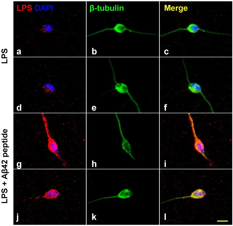
Increased affinity of LPS for neuronal nuclei in the presence of Aβ42 peptide. Panels (A–L) show LPS-neuronal interactions in the presence or absence of Aβ42 peptide; (A–F) in the absence of Aβ42 peptide and (G–L) in the presence of Aβ42 peptide. LPS preferentially associates with human neuronal nuclei both in Alzheimer’s disease (AD) and in LPS-addition experiments (Zhao et al., 2017a,b,c; Zhao and Lukiw, 2018a,b). Panels (A,D) show LPS (red) affinity for a polar region of a single DAPI-stained neuronal nucleus (blue). Panels (B,E) show single neuronal nucleus stained with neuron-specific β-tubulin III (green). Panels (C,F) show merged stain indicating LPS affinity for the polar region involving a single DAPI-stained neuronal nucleus. Panels (G,J) show the presence of Aβ42 significantly increases the affinity of LPS for single DAPI-stained neuronal nucleus. Panels (H,K) show single neuronal nucleus stained with neuron-specific β-tubulin III (green). Panels (I,L) show merged stain indicating LPS affinity for the neurite and soma of a single DAPI-stained neuronal nuclei. The results suggest that LPS is stimulated to associate with DAPI-stained neuronal nuclei in the presence of the hydrophobic Aβ42 peptide; neither Aβ40 peptide or β-actin showed comparable “association” effects (Zhao et al., 2017a,b,c; Lukiw et al., 2018); yellow scale bar (lower right) = 50 μm.

