Fig. 5.
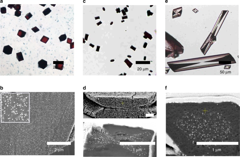
Calcite, gypsum and COM crystals precipitated in the presence of hydroxyl-NPs. a, b Calcite crystals precipitated in the presence of 0.05 wt% 14 nm PVA-NPs at [Ca2+] = 20 mM. c, d Calcium oxalate monohydrate (COM) crystals precipitated at [Ca2+] = 0.75 mM and [Ox] = 0.75 mM in the presence of 0.01 wt% 14 nm PGMA-NPs. e, f Calcium sulfate dihydrate (gypsum) crystals precipitated at [Ca2+] = 100 mM and [SO42−] = 200 mM in the presence of 0.08 wt% 14 nm PGMA-NPs. a, c, e show optical micrographs while b, d, f are SEM images of FIB cross-sections of the crystals.

