Fig. 8.
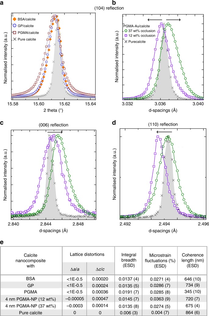
Powder XRD microstructure analysis of the protein/calcite and PGMA-NPs/calcite crystals. a {104} reflections of calcite crystals grown in the presence of proteins (BSA and GP) or PGMA, showing small shifts to larger d-spacings (lattice expansion) and peak broadening. b–d {104}, {006}, and {110} reflections obtained using high-resolution synchrotron powder XRD from calcite crystals with 12 wt% occlusion and 37 wt% occlusion. A shift to larger d-spacing is observed along the c-axis (expansion, seen in the {006} reflection (c)), and to a smaller d-spacing along the a-axis (contraction, as seen in the {110} reflection (d)). {104} reflections showed a transition from lattice expansion (12 wt% occlusion calcite) to contraction (37% occlusion calcite). e Summary of the strains and coherence lengths, where the lattice distortion and strain parameters were obtained using Rietveld analysis and line profile analysis, respectively.

