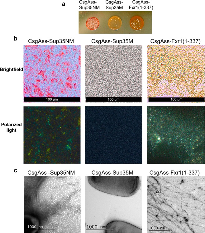
Figure 7.
FXR1 (1-337) fragment demonstrates amyloid properties in the bacteria-based C-DAG system. (a) E.coli cells producing CsgAss-Sup35NM and CsgAss-FXR1(1-337) proteins form red colonies whereas cells producing CsgAss-Sup35M form pale colonies on agar plates containing CR. (b) Micrographs of CsgAss-Sup35NM, CsgAss-Sup35M and CsgAss-FXR1(1-337) scraped cell samples harvested from CR-containing agar. Extracellular material containing CsgAss-Sup35NM and CsgAss-FXR1(1-337) binds CR (upper panel) and displays yellow-green birefringence when viewed between crossed polarizers (lower panel). Scale bar, 100 μM (c) Secreted CsgAss-Sup35NM and CsgAss-FXR1(1-337) proteins form fibrils visualized by transmission electron microscopy. Scale bar, 1000 nM. The E. coli cells are seen as dark spots on the middle image.

