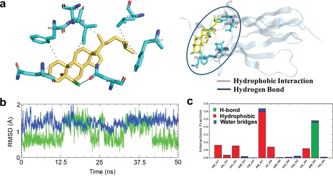
Figure 7.
Binding and interaction pattern of SCH at the fifth subdomain in the extracellular domain of TrkA receptor. Three-dimensional bound conformation of SCH in the TrkA, where it established direct interactions through hydrogen and hydrophobic bonds (a). Dotted lines represent the type of interactions between binding site residues and ligand. The results of MD simulation inferring as the RMSDs of protein and ligand that describe the overall conformational stability during 50 ns simulation (b). SCH mediated interactions to the binding site residues in TrkA receptor, categorized by hydrogen, hydrophobic and water bridge, respectively (c). Here each bar represents the percent of bond occupancy as a fraction.

