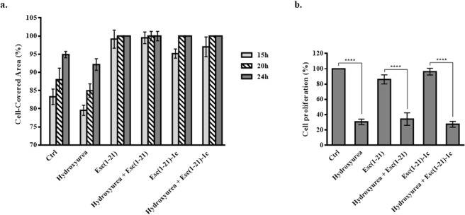
Figure 3.
Panel a: Quantitative assessment of cell migration in a pseudo-wound field produced in a monolayer of wt-CFBE cells. Cells were seeded and grown to confluence in each side of silicon chambers. After chamber removal, cells were treated with 250 μM hydroxyurea and 10 μM Esc(1–21) or 1 μM Esc(1–21)-1c. Some samples were incubated with each peptide or hydroxyurea alone, while cells incubated in culture medium were used as control (Ctrl). All experiments were performed four times in triplicates. Data are presented as percentages of cell-covered area at each time point. Percentages of samples treated with the combination hydroxyurea + peptide (with respect to samples treated with the peptide alone) were normalized to those previously obtained when CFBE were treated with each single peptide17. In parallel, percentages of samples treated with hydroxyurea alone (with respect to Ctrl) were normalized to those previously obtained for Ctrl samples17. All data are expressed as the mean ± standard errors of the mean (SEM). Panel b: Quantitative evaluation of cell proliferation after hydroxyurea treatment. Wt-CFBE cells (2 × 10⁴) were seeded in each well of a microtiter plate. After overnight incubation, cells were treated with 10 μM Esc(1–21), 1 μM Esc(1–21)-1c, 250 μM hydroxyurea or the combination of each peptide with hydroxyurea, for 24 h. After 2 h, cells were pulsed with BrdU. Cell proliferation was normalized to that of cells growing in culture medium (100% cell proliferation; Ctrl) and indicated as percentage. All data are the mean of three independent experiments ± SEM. Significance levels between groups is defined as P value of <0.0001 (****).

