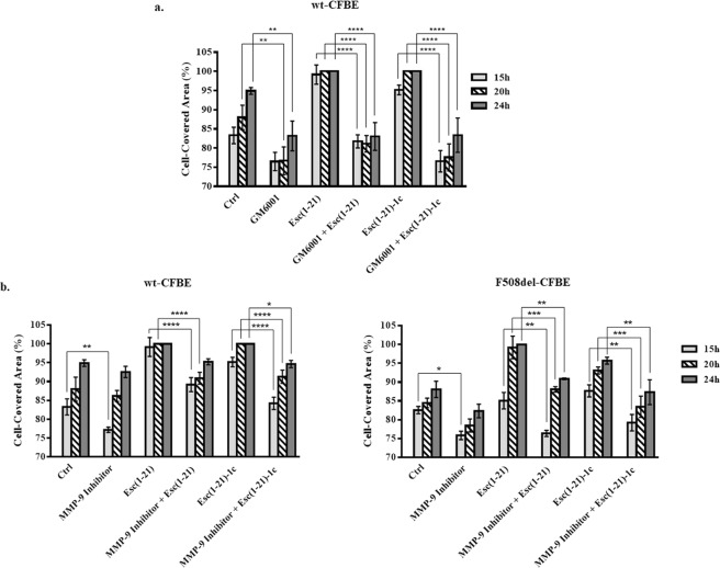
Figure 4.
Effect of GM6001 inhibitor (panel a) and MMP-9 inhibitor I (panel b) on the peptide-induced migration of CFBE cells. Before removing the silicone chamber, cells were pre-incubated for 30 min with GM6001 (25 μM) or with MMP-9 inhibitor I (20 nM and 50 nM for wt-CFBE and F508del-CFBE cells, respectively) and subsequently treated with 10 μM Esc(1–21) or 1 μM Esc(1–21)-1c + inhibitor. Some samples were treated with the peptide or inhibitor alone, at the same concentration used in the combination, while untreated cells were used as control (Ctrl). All experiments were repeated four times in triplicates. Cell-covered area was expressed as percentage at all time points. Percentages of samples treated with the combination inhibitor + peptide (with respect to samples treated with the peptide alone) were normalized to those previously obtained when CFBE were treated with each single peptide17. In parallel, percentages of samples treated with the inhibitor alone (with respect to Ctrl) were normalized to those previously obtained for Ctrl samples17. The results are the mean ± SEM. The levels of statistical significance among groups are P values of <0.05 (*), <0.01 (**), <0.001 (***) and <0.0001 (****).

