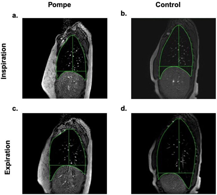
Figure 4.
Right sagittal thoracic MRI images at end-inspiration and end-expiration during an inspiratory capacity maneuver. At end-exhalation, lung volumes were similar in (a) subjects with late-onset Pompe disease (subject 3 shown) and in (b) healthy controls (subject 14 shown). (c) In Pompe disease, maximal inspiratory volume generation occurred exclusively through anterior-posterior (A-P) expansion of the chest wall. In contrast, (d) control subject generated volume primarily through diaphragm descent, measured as increased cranio-caudal (C-C) distance.

