Figure 2.

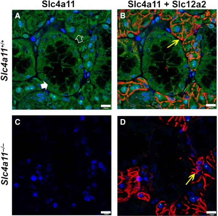
Immunolocalization of Slc4a11 in the mouse SMG. (A) Slc4a11 (green staining) expression in acinar (white open arrowhead) and duct cells (white filled arrowhead) of SMG in Slc4a11+/+ mice. (B) Merged image of Slc4a11 and basolateral Slc12a2 (red staining and yellow arrow, respectively) staining in Slc4a11+/+ mice. (C) Slc4a11‐specific immunostaining was not detected in the SMG of the Slc4a11 −/− mice. (D) Merged image of Slc4a11 and Slc12a2 (yellow arrow) staining from Slc4a11 −/− mice. Images shown are from male Slc4a11+/+ and Slc4a11 −/− mice. Scale bar = 10 μm; nuclei were stained with DAPI (blue).
