
Figure 2. ΔNC16A mice show significantly increased melanoma progression in both flank and ear injection models.
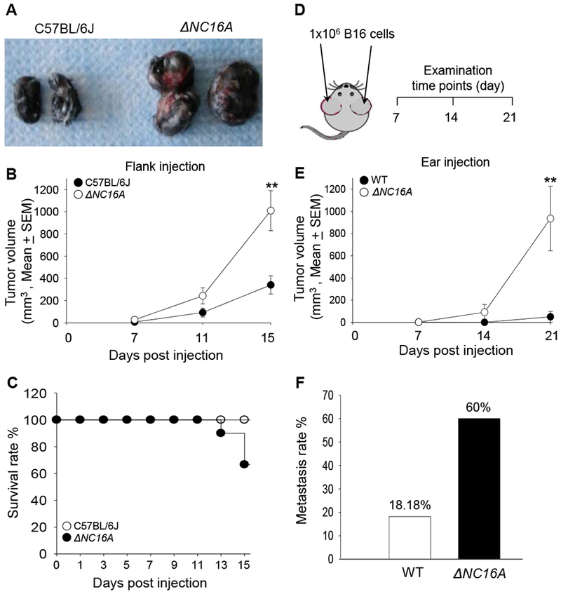
Eight weeks old of C57BL/6J and ΔNC16A mice were injected at the flank (A-C) or ear (D-F) with 1×10⁵ or 1×10⁶ B16 melanoma cells, respectively, and examined at different time points post injection. In the flank model, ΔNC16A mice developed significantly larger size of tumors (A), significantly increased tumor growth rate starting from day 11 (B) (** p<0.05, Scheirer-Ray-Hare test; n=9, for both WT and ΔNC16A mice.) and reduced survival rate at day 15 post injection (C).Using the cox regression model, we obtain an expected 2.55 fold (95% CI 0.66-9.89) the hazard ratio of NC16A comparing to the wild-type group, with p-value of 0.156 in the likelihood-ratio test. In the ear model (D), ΔNC16A mice showed significantly faster tumor growth starting day 14 (E) (** p<0.01, Scheirer-Ray-Hare test) and significantly increased rate of developing neck lymphatic metastasis at day 21 post melanoma cell injection (p<0.05, Fisher’s Exact Test) (F) For ear injection mode, n=10 for ΔNC16A, n=22 for WT).