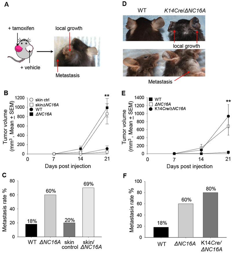
Figure 3. Skin-and basal keratinocyte-specific BP180 dysfunction are sufficient to promote B16 melanoma progression.

A, TamCre-NC16A mice were treated at the left ear with tamoxifen (red) and right ear with vehicle control (white). B16 melanoma cells (1× 10⁶cells) were injected into both tamoxifen-and vehicle-treated ears 10 days post tamoxifen treatment. At 21 days post melanoma injection, only the tamoxifen-treated ear developed local tumor and lymphatic metastasis. B, Tumors volumes in tamoxifen-treated ears were significantly increased starting from day 14 post melanoma cell injection compared to tamoxifen-treated WT ears (skin control) (** p<0.01, Scheirer-Ray-Hare test; n=13 for skinΔNC16A, n=15 for skin control). C, A significantly larger proportion (69%) of tamoxifen-treated TamCre-NC16A mouse (skinΔNC16A) ears also developed lymphatic metastasis as compared to 20% of skin control at 21 days post injection. (p<0.05, Fisher’s Exact Test; n=13 for skinΔNC16A, n=15 for skin control). D-F, Ears of 8 weeks old basal keratinocyte-specific ΔNC16A (K14Cre/∆NC16A) mice were injected with B16 melanoma cells (1×10⁶ cells) and monitored for 21 days. K14Cre/∆NC16A mice developed significantly larger tumors at day 21 (D), and the significantly increased tumor growth started at day 14 (** p<0.01, Scheirer-Ray-Hare test; n=8 for K14Cre/∆NC16A mice, n=22 for WT) (E). In addition, 80% of K14Cre/∆NC16A mice developed lymphatic metastasis as compared to 18% of WT mice at day 21 post melanoma injection (Fisher’s Exact Test, p<0.01 n=8 for K14Cre/∆NC16A mice, n=22 for WT) (F).
