Fig 5:

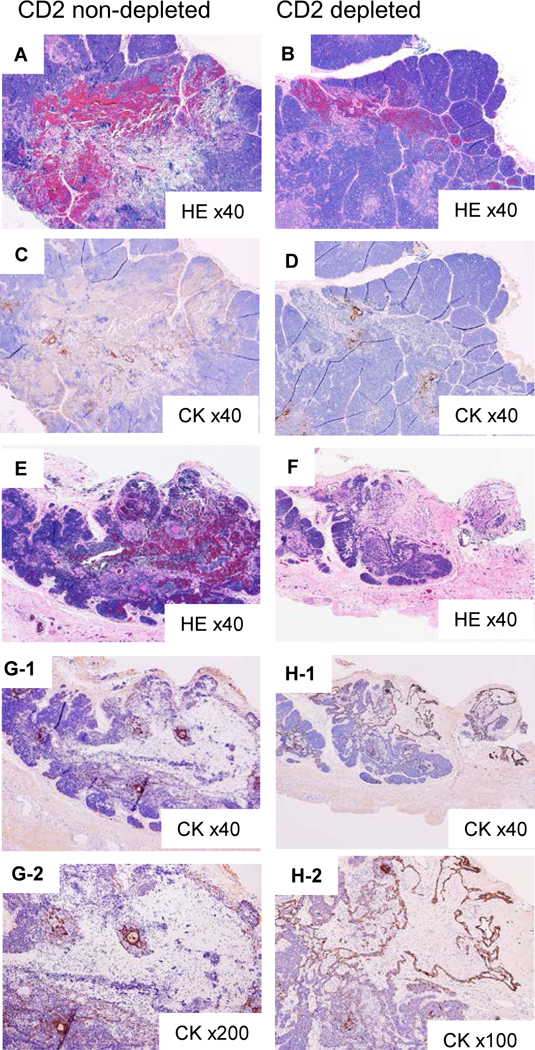
Histologic findings of injected sites of cynomolgus monkeythymic cells in porcine cervical thymic lobes at POD 16 in Group 2 (#28090), demonstrating rejected cynomolgus monkey TEC in the porcine thymic lobe. A, B) HE staining of CD2 non depleted and CD2 depleted thymic cells in #28090 (x40). C, D) CK staining of the same fields of A and B (x40). E, F) HE staining of CD2 non depleted and CD2 depleted thymic cells in #28091 (x40). G-1 shows CK staining of the same fields of E (x40), and G-2 shows a high-power view (x200). H-1 shows CK staining of the same fields of F (x40), and H-2 shows a high-power view (x200).
