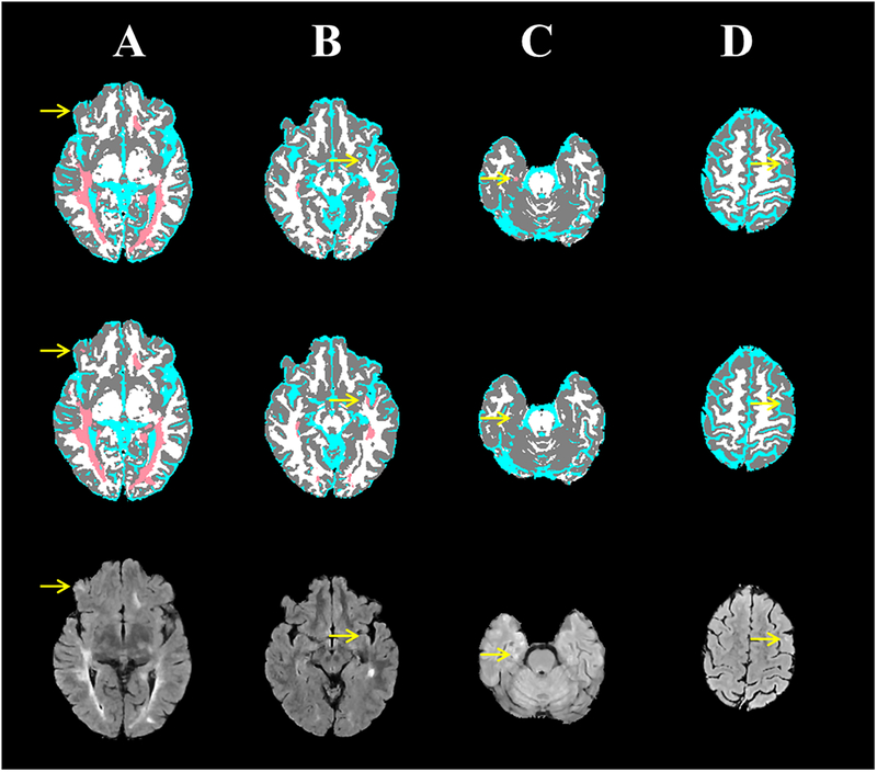
Figure 6:
Four MS cases (columns) showing the brain segmentation from the expert-validated ground truth (top row) and the CNN (middle row), and corresponding FLAIR images (bottom row). (A) A small subcortical lesion that was detected by the CNN, but was missed in the “ground truth”. (B) A slight hyperintensity in subcortical white matter was mistakenly segmented by the CNN as a T2 lesion. (C) A mediotemporal lesion that was not detected with CNN. (D) A subcortical lesion that was missed by CNN.

