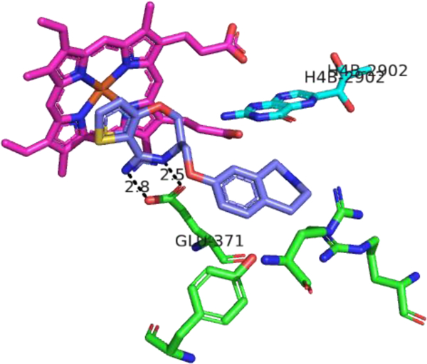
FIGURE 8.

X-ray crystal structure of compound 21 (purple) bound in the murine iNOS active site (PDB ID: 3EBF166. Heme is depicted in magenta, H4B in cyan, and amino acid residues of the enzyme in green. Distances are measured in Ångstroms. Figure prepared using PyMol (www.pymol.org).
