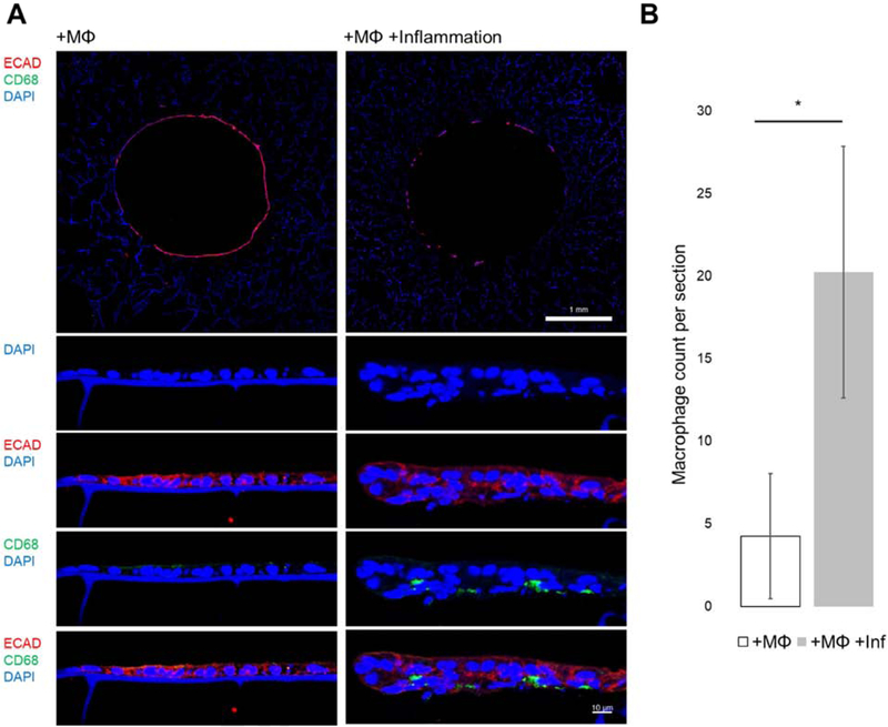
Figure 4. Monocyte-derived macrophages infiltrating the epithelial monolayer under inflammation.
(A) Transverse histological sections of models at day 3 show e-cadherin positive epithelial cells and little to no CD68+ cells at 10X (top scalebar 1 mm). Higher magnification images of the epithelium show CD68+ macrophages on the basal side of the epithelium of inflamed conditions (bottom scalebar 10 μm). (B) Quantification of CD68+ macrophage cells on transverse sections between +MO and +MO+Inflammation show statistical significance (n=4) (p<0.05).

