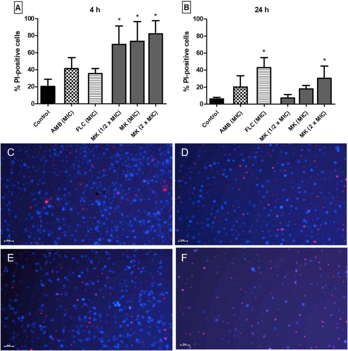
Figure 1.
Effect of different concentrations of MK58911 (MK – 1/2 × MIC; MIC and 2 × MIC), amphotericin B (AMB–MIC), or fluconazole (FLC–MIC) on C. neoformans membrane, evaluated by percentage of propidium iodide (PI)-positive cells at (A) 4 h and (B) 24 h. *p < 0.05 compared to control group. Representative images of C. neoformans (C) non-treated cells or (D) cells treated at MIC of amphotericin B (E) MIC of fluconazole or (F) MIC of MK58911 at 4 h. The cell wall of all fungal cells was stained with calcofluor (blue), and cells with membrane damage were stained with propidium iodide (red). Scale bar: 20 μm.

