Figure 4.

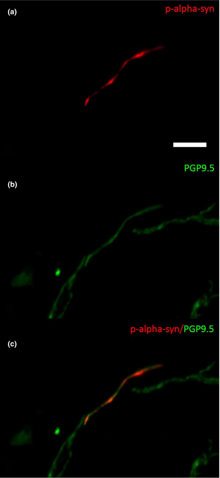
Photomicrograph of a double‐immunofluorescence staining with anti‐α‐synuclein (a, c) and anti‐PGP9.5 (b and c) in a biopsy of a patient with PD. P‐α‐synuclein is detectable within a dermal nerve fiber (c). Bar = 20 µm

Photomicrograph of a double‐immunofluorescence staining with anti‐α‐synuclein (a, c) and anti‐PGP9.5 (b and c) in a biopsy of a patient with PD. P‐α‐synuclein is detectable within a dermal nerve fiber (c). Bar = 20 µm