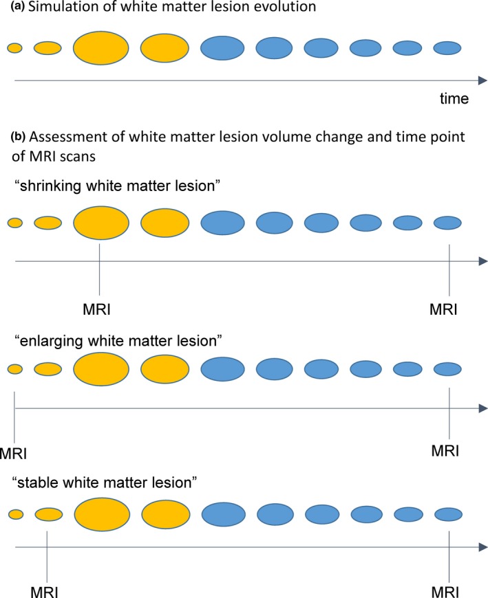
Figure 1.

(a) A conceptual scheme of white matter lesion evolution is shown. (b) By three scenarios the dependency of the assessment of white matter lesion volume change on the time point of MRI scans is illustrated. The time points of MRI scans are indicated by a vertical black line. Gadolinium enhancement is illustrated by yellow coloring. Timing of the initial MRI scan may decide whether analysis of the same WML after 1 year demonstrates shrinkage (b, top panel), enlargement (b, middle panel), or stability (b, bottom panel)
