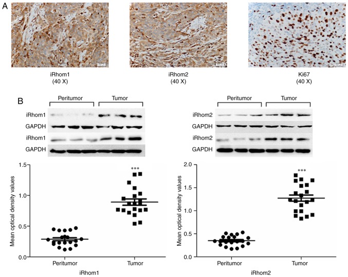
Figure 1.
Expression of iRhom1, iRhom2 and Ki-67 in cancerous tissues and adjacent normal tissues of patients with CC. (A) Representative photomicrographs (40X) of CC specimens stained with anti-iRhom1, anti-iRhom2 and anti-Ki67 antibodies. iRhom1 and iRhom2 were in the cytoplasm, and Ki-67 was mainly in the nucleus, as indicated by dark-yellow granules. (B) Quantitation of results, revealing increased levels of iRhom1 and iRhom2 in cancerous tissues relative to paired adjacent non-cancerous tissues (***P<0.001). iRhom1, inactive rhomboid protein 1; iRhom2, inactive rhomboid protein 2; CC, cervical cancer.

