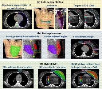
Figure 1.

Auto planning workflow. (a) Auto‐segmentation: normal structures including bilateral lungs, heart, and spinal cord were contoured using atlas‐based segmentation; the superior, inferior, lateral and medical boundary landmarks were identified from the “box” and “border” contours or the wires placed on the patient skin. Two chest wall contours were identified from the computed tomography; targets were segmented following RTOG 1005 recommendations. (b) Beam placement: two tangent beams were placed based on the boundary points and chest wall points. The gantry angle, collimator angle, and jaw positions were then optimized to maximize target coverage and minimize normal lung and heart volume in the beam. Beam energy was selected based on the maximal separation; (c) Hybrid IMRT: the automatic breast plan includes two prescriptions: a three‐dimensional (3D) prescription with two static tangent beams and an intensity modulated radiation therapy (IMRT) prescription with two step‐and‐shoot tangent beams. The beam weightings of the 3D prescription were optimized using dose points selected uniformly inside PTVeval_breast. The 3D prescription delivers full prescription dose to the maximum dose. The IMRT prescription was optimized to deliver uniform dose to breast and reduce dose to lungs and heart.
