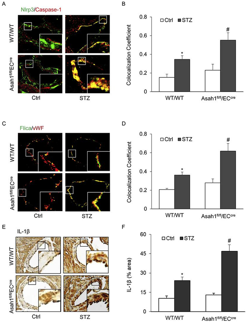
Fig. 1. AC deficiency increased the formation and activation of NLRP3 inflammasomes in the coronary arterial endothelium of STZ-treated mice.

Wild type (WT/WT) and Asah1 endothelial specific knock-out mice (Asah1fl/fl/ECcre) were treated with vehicle (Ctrl) or streptozotocin (STZ) as described. A. Representative fluorescent confocal microscopic images displaying the yellow dots or patches showing the colocalization of NLRP3 (green) with caspase-1 (Red). B. The summarized data showing the colocalization coefficient of NLRP3 with caspase-1. C. Representative fluorescent confocal microscopic images displaying the yellow dots or patches showing the colocalization of a green fluorescent probe specific for active caspase-1, FLIC A (green) with vWF (Red), an endothelium marker. D. The summarized data showing the colocalization coefficient of FLIC A with vWF. E. Representative immunohistochemical staining showing IL-1β accumulation in the coronary arterial wall. F. The summarized data showing the density of IL-1β stained with selective anti-IL-1β antibody. Data are expressed as means ± SEM, n=5. * p<0.05 vs. WT/WT-Ctrl group; # p<0.05 vs. WT/WT-STZ.
