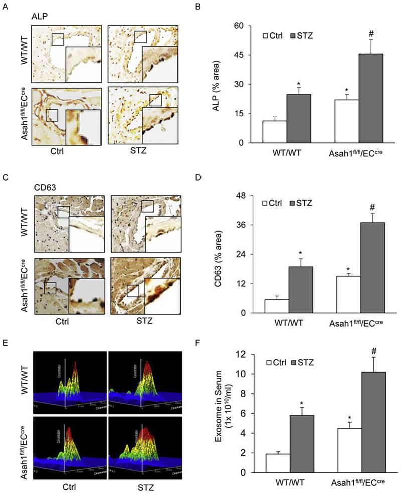
Fig. 3. AC deficiency enhances exosome secretion in the coronary arterial endothelium of STZ-treated mice.

A. Representative microscopic images of tissue slide with IHC staining showing the expression of exosome marker CD63 in the mouse coronary arterial wall. B. The summarized data showing the density of CD63 stained with the anti-CD63 antibody. C. Representative microscopic images of tissue slide with IHC staining showing the expression of exosome marker ALP in the mouse coronary arterial wall. D. The summarized data showing the density of ALP stained with the anti-ALP antibody. E. Representative 3D histograms showing the secretion of exosomes in the mouse serum as measured by nanoparticle tracking analysis (NTA) using NanoSight NS300 nanoparticle analyzer. F. The summarized data showing the released exosomes in the serum. Data are expressed as means ± SEM, n=5. * p<0.05 vs. WT/WT-Ctrl group; # p<0.05 vs. WT/WT-STZ.
