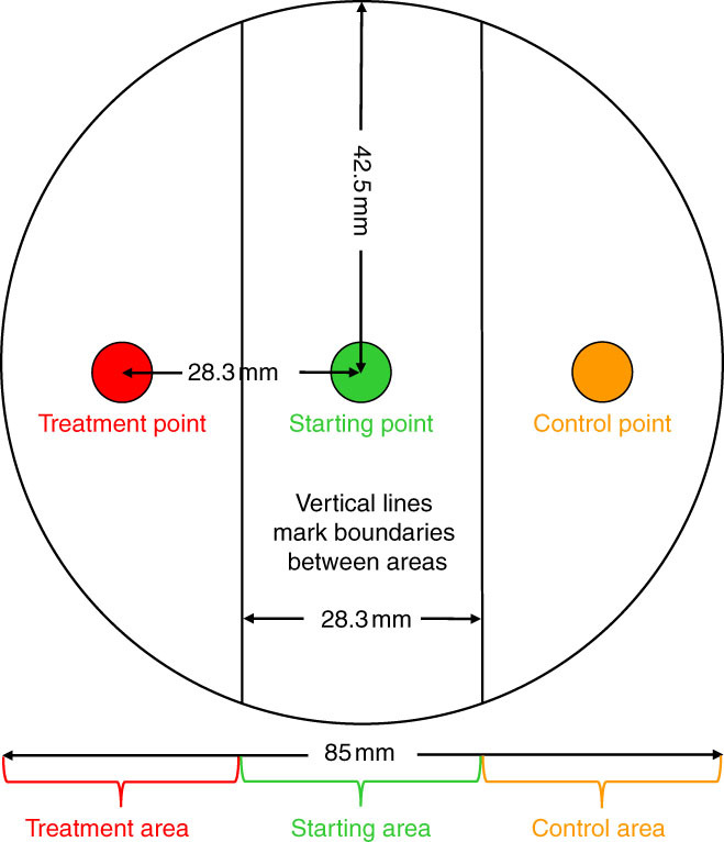
Figure 1.

Diagram of a chemotaxis assay. Each plastic plate was divided into three areas: the treatment area (consisting of the treatment point and the surrounding area), the starting area (consisting of the starting point and the surrounding area), and the control area (consisting of the control point and the surrounding area). J2 were placed on the agar at the starting point, treatments were applied to the agar at the treatment point, and sterile distilled water was applied to the agar at the control point. Measurements are given for a plate with an 85-mm-diam. cup (100-mm-diam. lid).
