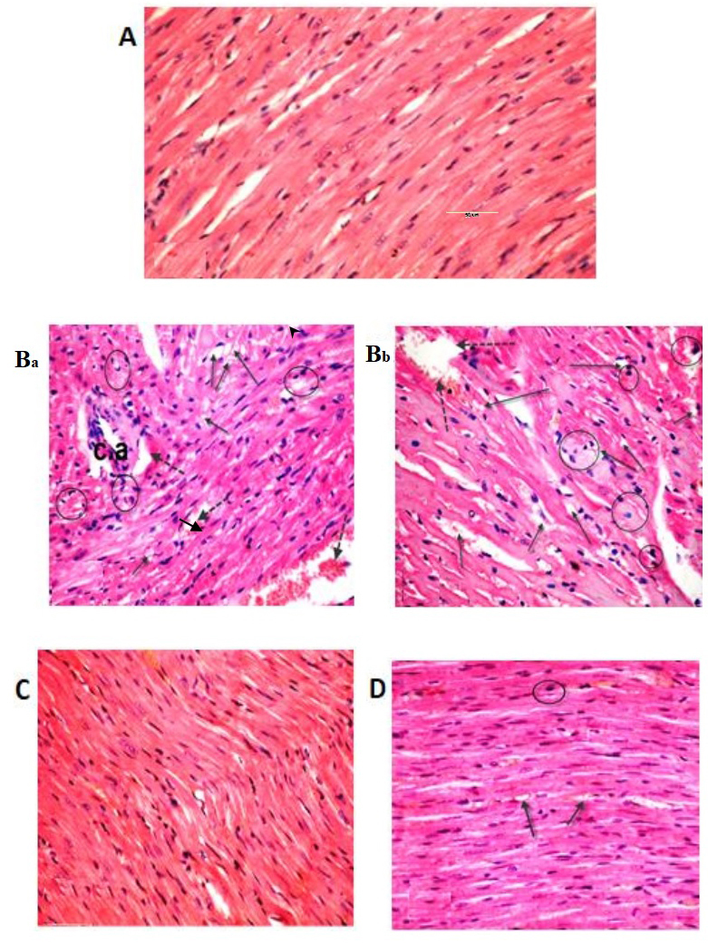
Figure 5.

Light micrographs of heart tissue. The (A) control and sesame lignans-treated rats (C) exhibited normal myocardial fiber architecture, with branching muscle fibers and centrally located oval nuclei. (Ba and Bb) Sections of heart tissue treated with BPA exhibited a loss of normal architecture, widespread fragmentation and muscle fiber degeneration. Inflammatory and mast cells (black circle) were present in the connective tissue, along with a congested blood vessel (dotted arrow), vacuoles (black arrow) and branches of thickened coronary vessels. (D) Histological alterations induced after BPA-treatment were markedly reduced when treated with BPA and sesame lignans (H&E staining; magnification, ×400). BPA, bisphenol A; c.a. coronary artery.
