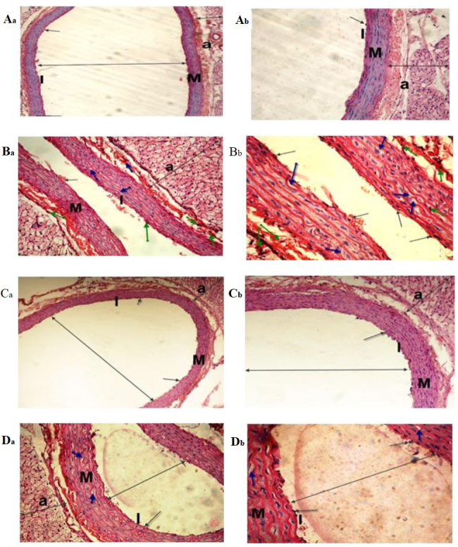
Figure 6.

Light micrographs of the dorsal aorta. The (Aa and Ab) control and (Ca and Cb) sesame lignans-treated group exhibited normal tunica intima with a single irregular layer of endothelial cells (black arrow). The tunica media, comprising elastic fibers and the tunica adventitia also appeared normal. (Ba and Bb) The BPA-treated group exhibited sclerotic changes in the walls and atrophy of elastic fibers (green arrows), loss of the tunica intima endothelial cells, disorganized and vacuolation of the tunica media (blue-arrow), increased adventitial thickness and decreased lumen size of the aorta (double-headed arrows) compared with the control group. (Da and Db) Histological changes induced after BPA-treatment were markedly reduced in the BPA and sesame lignans-treated group (H&E staining; Aa, Ba, Ca and Da magnification, ×200; Ab, Bb, Cb, and Db magnification, ×400). I, tunica intima; M, tunica media; a, tunica adventitia.
