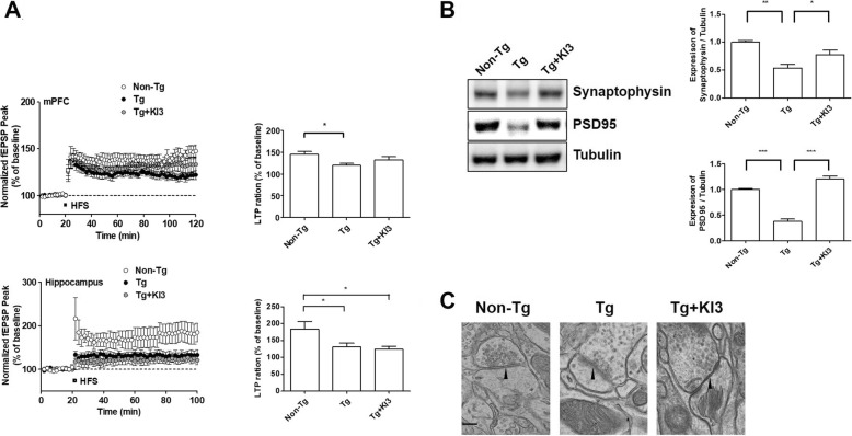
Fig. 5.
EA increases synaptic plasticity and reduces synaptic ultrastructural degradation in 5XFAD mice. a EA treatment increases long-term potentiation (LTP) in the prefrontal cortex and hippocampus of 5XFAD mice. The peaks of field excitatory synaptic potentials (fEPSPs) were normalized using baseline recordings (n = 6/group; a, left). The graph shows cumulative data displaying the average fEPSP of 90–100 min in the prefrontal cortex and hippocampus after EA treatment (n = 6/group; a, right). b Representative images of immunoblots displaying increased synaptic marker protein levels (synaptophysin and PSD-95) after EA treatment in 5XFAD mice. Tubulin was used as the loading control. Quantitative analysis of the expression levels of synaptophysin and PSD-95 (n = 3–4/group). c Representative transmission electron microscopy images of synaptic structures (pre- and postsynapse, synaptic space, and synaptic vesicles). Black arrowheads indicate the clearly visible synaptic cleft. Scale bar, 200 μm. Data are presented as means ± SEM (*p < 0.05, **p < 0.01, ***p < 0.001)

