Fig. 1.
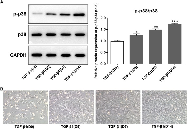
Expression of p-p38/p38 in TGF-β1-induced BMSCs and morphological observation. a Western blotting of p-p38 and p38 expression in BMSCs following TGF-β1-induced for 0, 5, 7, and 14 days. Relative protein expression of p-p38/p38 was elevated on a time-dependent manner under the TGF-β1 induction when compared with day 0. b Representative images of BMSCs. Morphological changes of BMSCs following TGF-β1 induced for 0, 5, 7, and 14 days were observed under an inverted microscope. On day 0, BMSCs grew adherently to the wall, and the cells were triangular or polygonal in shape. From day 5 to day 14, cell morphology changed significantly and gradually presented a typical paving stone shape with uniform size and shape. *p < 0.05, **p < 0.01, and ***p < 0.001 vs. TGF-β1 (D0)

