Fig. 1.
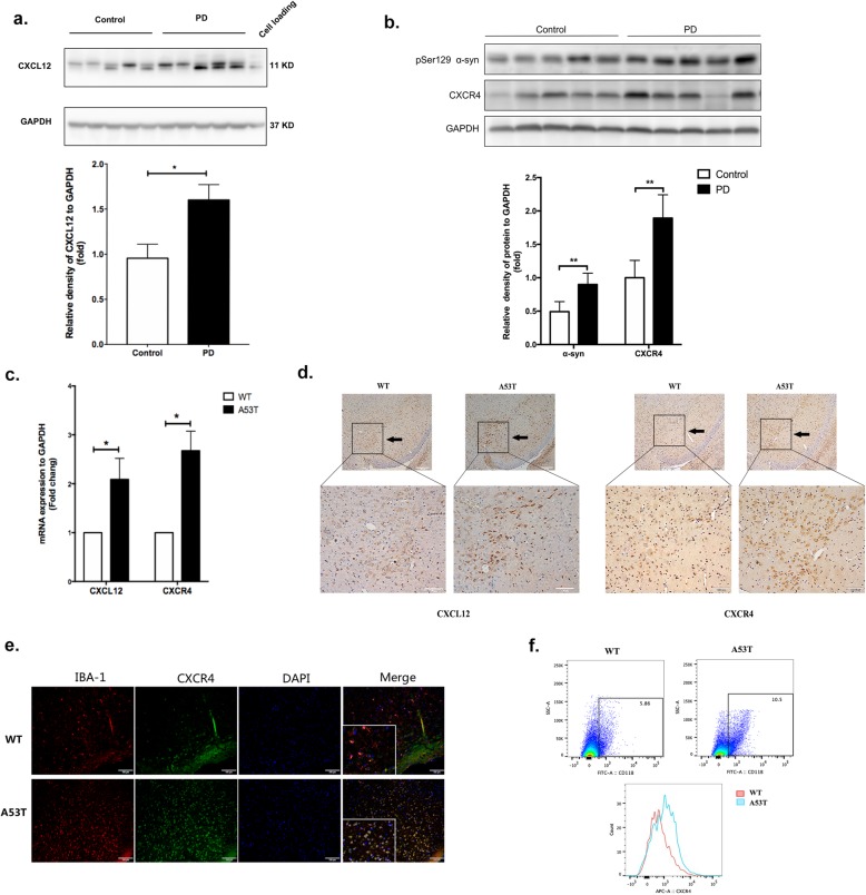
The levels of CXCL12 and CXCR4 were elevated in the brain tissues of PD patients and A53T transgenic mice. a Western blot analysis showed the expression of CXCL12 in SN sections of postmortem brain tissue from control and PD subjects. GAPDH was used as an internal control for gel loading, and M17 cell protein was loaded as a positive control. b Western blot analysis showed an upregulation of CXCR4 and pser129 a-syn protein levels in human PD brains compared with the control. c The mRNA expression of CXCL12 and CXCR4 was determined by real-time qPCR in A53T and WT mice. n = 3 per group. d Brain slices from the substantia nigra were stained for CXCL12 and CXCR4 by immunohistochemistry. n = 3 per group for A53T and WT mice. e Brain slices from the substantia nigra were double stained for IBA-1 (red) and CXCR4 (green). Images were captured with a fluorescence microscope. Scale bar = 100 μm. n = 3 per group. f Brain tissue from the substantia nigra was stained for microglia (CD11b-FITC) and CXCR4 (APC), and the frequency of the CD11b+ and CD11b+CXCR4+ cells was assessed by flow cytometry. n = 3 per group for A53T and WT mice. The data are shown as the mean ± SEM for three independent experiments. **p < 0.01. *p < 0.05

