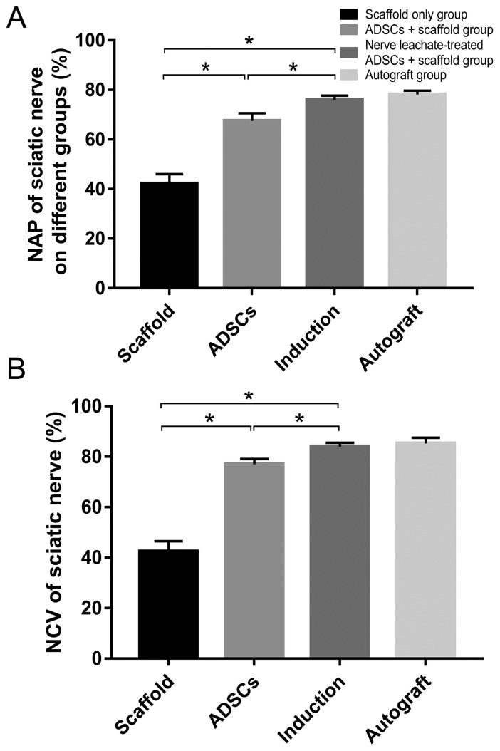
Figure 3.
Electrophysiological assessment. (A) Electrical stimuli were applied to the proximal portion of the nerve trunk, and the amplitude of the NAP was recorded. (B) NCV was analyzed as a function of passing distance and time. The data are presented as the mean ± SD. *P<0.05. ADSC, adipose-derived mesenchymal stem cell; NAP, nerve action potential; NCV, nerve conduction velocity.

