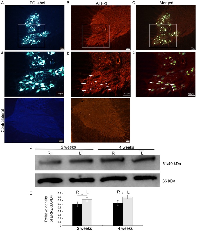
Figure 1.
FG labeling and ATF-3/ERRγ in the injured spinal cord. FG retrograde labeling was performed in the representative axotomy side C7 and C8 spinal cord sections examined under the fluorescence microscope at day 3. (A) FG localization in the cell somata and cell processes of neurons whose axons were cut at the level of the brachial plexus roots. Scale bar=100 µm, magnification ×10. (a) Magnification (×20) of the area marked in image A. Scale bar=100 µm. (B) AFT-3 nuclei staining on the axotomized side of the C7 and C8 spinal cord sections examined under the fluorescence microscope at day 3. No staining was observed on the contralateral side (not in view). Magnification ×10 (b). Magnification (×20) of the area marked in image B. Scale bar=100 µm. (C) A merged image demonstrating co-localization of FG and ATF-3 in injured (axotomy) motor neurons at day 3. Magnification ×10. (c) Magnification (×20) of the area marked in image C. Scale bar=100 µm. (D) A representative western blot analysis gel. The ERRγ/GAPDH ratio for the L (uninjured side) and R (injured side) halves of the spinal cord of each at 2 and 4 weeks post-avulsion. (E) Densitometry results are presented as means ± standard error of the mean. The level of ERR γ protein was significantly decreased on the right side (brachial plexus injured) of the spinal cord at both time points. *P<0.05. The black bars represent the protein level on the injured side, and the gray bars represent the expression levels on the contralateral, uninjured side. FG, Fluorogold; ATF-3, cyclic AMP-dependent transcription factor 3; ERRγ, estrogen-related receptor γ; L, left, contralateral side; R, right ipsilateral side.

