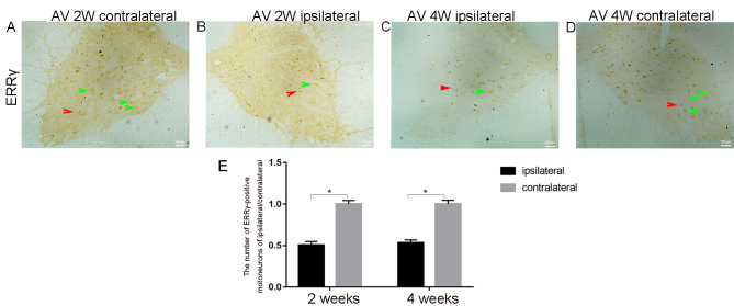
Figure 3.
Representative micrographs used for the counting of the ERRγ positive motor neurons on both sides of the same C7 segment of the spinal cord. (A) The contralateral side at 2 weeks. (B) The ipsilateral side at 2 weeks. (C) The ipsilateral side at 4 weeks. (D) The contralateral side at 4 weeks; all at magnification ×20) In the ERRγ-stained sections, the numbers of positive motor neurons were notably decreased on the ipsilateral side compared with those on the contralateral side. Red arrows show positive ERR γ signals in smaller γ motor neurons; Green arrows depict positive ERR γ signals in large α motor neurons. (E) The mean of the total number of ERRγ-positive motor neurons in 10 serial sections of both ventral horns of C7 spinal segments were computed at 2 and 4 weeks post-avulsion. The average number of ERRγ-positive motor cells on the injured side (black column, ipsilateral) were significantly decreased compared with that identified on the contralateral side (gray column, contralateral). *P<0.05. ERRγ, estrogen-related receptor γ.

