. 2019 Dec 17;45(6):1129–1135. doi: 10.1590/S1677-5538.IBJU.2019.0085

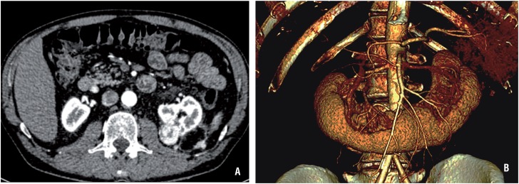
Figure 1. A) Angio-TC scan showing a 3.3 x 2.6 cm tumor in previous surgical bed, partly exophytic and reaching the renal sinus. Three solid nodules in the left posterior pararenal fascia of 0.9, 1.3 and 1.6 cm are also identified. There are identified two main renal arteries, one left and one right, and three accessory arteries originated in the anterior margin of the aorta, directed behind the renal isthmus, two to the left and one to the right. Besides, there are two left renal veins, the inferior with a retroaortic trajectory, and three right renal veins. B) Picture of the 3D reconstructed renovascular video images.

Figure 1